Brillouin
Spectroscopy
What is
Brillouin
Spectroscopy?
All-optical Brillouin spectroscopy is a non-contact and label-free technique used to explore the mechanical properties of materials at microscopic scales.
In Brillouin spectroscopy, light interacts with intrinsic acoustic waves of materials, so that a small portion of the scattered light forms tiny spectral peaks. By measuring the frequency shift and linewidth of the Brillouin peaks, one can determine the elastic moduli of the material analyzed, providing fundamental information about the material related to the stiffness and the viscosity.
When applied to biological samples, such as cells and tissues, this technique offers unique capabilities in analyzing their associated biomechanics non-destructively and at micron-scale spatial resolution in 3D. As such, Brillouin spectroscopy holds great potential to be employed as an essential tool in the biomedical domain, where the biomechanical changes are responsible for the onset and progression of several diseases.
How it all
started?
Fun fact
Around the same time as Léon Brillouin, Soviet physicist Leonid Mandelstam is believed to have recognised the possibility of this scattering process in 1918, but he did not publish his findings until 1926. To give credit to Mandelstam, the effect is sometimes referred to as Mandelstam-Brillouin scattering.
Brillouin spectroscopy began with the brilliant work of French physicist Léon Brillouin, who first explained how light interacts with acoustic waves in a material back in 1922. Léon was one of the pioneering physicists who lived in the early 20th century, and he is considered one of the founders of modern solid-state physics.

1927 Solvay conference participants. L. Brillouin at the top right stands just above M. Born and N. Bohr and next to W. Pauli, W. Heisenberg, and R.H. Fowler.
He predicted the interaction between optical waves (photons) and acoustic waves (phonons), where a photon interacts with density fluctuations in matter resulting in a negative (Stokes) and a positive (anti-Stokes) frequency shift, as shown schematically below:

A schematic of the Brillouin spectrum
Brillouin Spectroscopy: A history lesson
The phenomenon of Brillouin light scattering, named after Léon Brillouin, was first described in 1922 when he detailed the interaction between optical waves and acoustic waves within a medium.
Therefore the scientific community refers to the phenomenon as “Brillouin scattering” or “stimulated Brillouin scattering (SBS),” depending on whether the scattering occurs spontaneously or through external stimulation.
Key phases of Brillouin Spectroscopy

(Read more)
(Read more)
(Read more)
(Read more)
(Read more)
(Read more)
(Read more)
(Read more)
(Read more)
(Read more)
More info
What does it measure?
In Brillouin spectroscopy, light exchanges energy with intrinsic acoustic waves of materials, so that a small portion of the scattered light undergoes tiny frequency shifts, resulting in a Stokes and an anti-Stokes Brillouin peak. A measure of the Brillouin frequency shift \(\nu_{B}\) and linewidth \(\Delta\nu_{B}\) provides information about the viscoelastic properties, and in particular on the elastic moduli forming the full elastic tensor of a material.
In the backscattering geometry commonly used in confocal microscopy, Brillouin spectroscopy determines the complex longitudinal modulus M(ν) = M‘ (ν) + iM”(ν).
The real part of M, also known as the storage modulus M‘, reflects the material’s elastic behavior, or the energy stored within the material. On the other hand, the imaginary part of the modulus, also known as the loss modulus M”, reflects a material’s acoustic attenuation and its viscosity-like properties.
How does it work?
Brillouin spectroscopy functions similarly to other spectroscopy techniques, such as Raman spectroscopy. However, detecting the Brillouin spectrum poses more severe challenges. The Brillouin frequency shift, typically less than \(30\,\mathrm{GHz}\) (\(1\,\mathrm{cm}^{-1}\)) in biological samples, occurs very close to the pump laser frequency and is almost indistinguishable from the Rayleigh scattered light. Compounding this difficulty is the fact that Brillouin scattering is typically more than \(10^{9}\)-fold weaker than Rayleigh scattering.
These challenges impose the need for critical components. First, a laser with a narrow linewidth and low amplified spontaneous emission (ASE) is essential to minimize noise. Unlike Raman spectroscopy, where the signal is well-separated (typically \(>1\,000\,\mathrm{cm}^{-1}\)) from the Rayleigh scattered light, Brillouin detection further requires ultra-narrow and high-rejection filters capable of suppressing the elastic background light. Additionally, a spectrometer with sub-\(\mathrm{GHz}\) resolution is needed to accurately resolve the Brillouin peaks. These requirements typically lead to complex, low-throughput and bulk instrumentation.
Specto's solutions
At Specto, we aim to make Brillouin spectroscopy accessible to everyone interested in measuring micromechanical properties.
Our groundbreaking solutions help research users to easily acquire relevant spectral data, making Brillouin spectroscopy more accessible and adaptable to a wide range of biomedical and industrial applications.
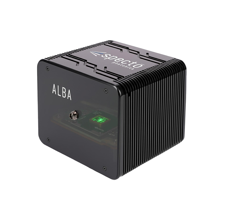
ALBA:
ON-CHIP BRILLOUIN SPECTROMETER
We are developing groundbreaking solutions, such as on-chip Brillouin spectrometers, leveraging photonic integrated circuits, that provides extreme miniaturization, ease of use and portability. This technology permits eliminating the need of aligning optical parts and providing superior stability and compactness.

BIPD:
HIGH-REJECTION FILTER
Additionally, our proprietary high-rejection BIPD optical filter enhances the detection of Brillouin spectra, even in highly turbid materials. This Specto’s proprietary technology uses birefringent crystals to effectively reject the unwanted Rayleigh scattering with an extinction ratio of by >60 dB.